






Diseases & Conditions
Shoulder Joint Labral Tear
This article was written and/or reviewed by a member of American Shoulder and Elbow Surgeons (ASES).
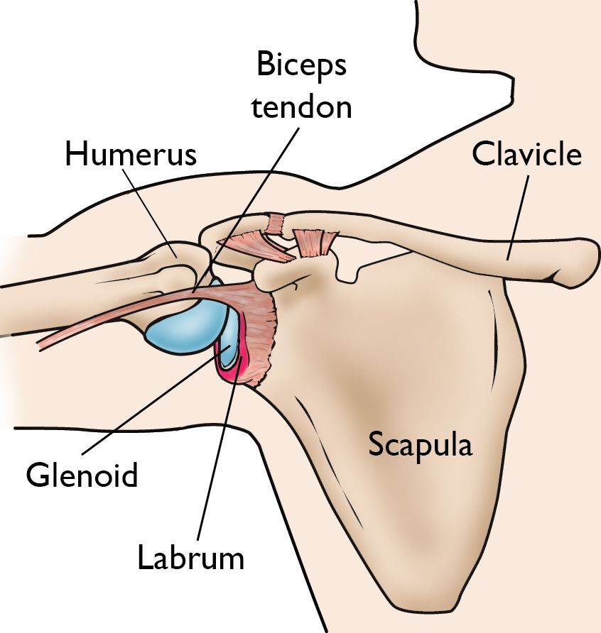
The labrum is a rim of cartilage tissue in your shoulder that helps deepen the shoulder socket. It also provides an attachment point for ligaments that help stabilize the shoulder and for one of the tendons of the biceps muscle.
If you tear the labrum, it can result in deep pain in the shoulder with many different motions and can sometimes lead to feelings of instability or dislocations of the joint.
Traumatic labral tears often occur when a person suffers a shoulder dislocation. Other tears, especially tears near where the biceps tendon attach, are seen in athletes due to repetitive stress on the tendon. Labral tears can also develop due to normal wear and tear of the joint, along with the development of arthritis.
While not all of these tears need to be treated with surgery, tears that cause ongoing pain or chronic instability can be treated surgically with good results.
Anatomy of the Shoulder
The shoulder is a ball-and-socket joint made up of three bones:
- The humerus (upper arm bone)
- The scapula (shoulder blade)
- The clavicle (collarbone)
The head of the upper arm bone (humeral head) rests in a shallow socket on the shoulder blade called the glenoid. The head of the upper arm bone is much larger than the socket, and the labrum surrounds the socket to help stabilize the joint. The labrum deepens the socket by up to 50%. It also serves as an attachment site for several ligaments that help keep the shoulder stable and for a tendon of the biceps muscle.
Types of Labral Tears
There are several types of labral tears:
- A SLAP lesion (superior labrum, anterior [front] to posterior [back]) is a tear of the labrum that usually occurs on the upper part of the socket and may also involve the origin, or starting point, of the long head of the biceps tendon. This type of tear is often seen in athletes due to repetitive stress on the biceps tendon.
- A tear of the anterior (front) part of the labrum at the bottom of the socket is called a Bankart lesion. This usually happens from an anterior shoulder dislocation (a dislocation when the humeral head comes out of the front of the socket).
- A tear of the labrum can also occur in the back part of the socket. This is called a posterior labral tear. This type of tear can be a traumatic tear due to injury. It can also develop due to normal wear and tear on the shoulder, also known as osteoarthritis.
What Can Cause a Labral Tear?
Injuries to the tissue rim surrounding the shoulder socket can occur from acute trauma or repetitive shoulder motion. Examples of traumatic injury include:
- Falling on an outstretched arm
- A direct blow to the shoulder
- A sudden pull, such as when trying to lift a heavy object
- A violent overhead reach, such as when trying to stop a fall or slide
Injuries to the labrum from repetitive shoulder motion are often seen in throwing athletes or weightlifters.
Symptoms of a Labral Tear
The symptoms of a labral tear can be very similar to those of other shoulder injuries and are not always specific. Symptom also typically depend on which part of the labrum is torn.
Symptoms can include one or more of the following:
- A sense of instability in the shoulder
- Shoulder dislocations
- Deep shoulder pain, usually with overhead or reaching activities
- Catching, locking, popping, or grinding in the shoulder
- Occasional night pain or pain with daily activities
- Decreased range of motion
- Loss of strength
Diagnosing a Labral Tear
Medical History and Physical Examination
If you are experiencing pain or instability of your shoulder, your doctor will likely ask whether there was a specific injury that caused your symptoms or if the symptoms came on gradually. They will ask you to describe your symptoms, including which activities typically cause them.
The doctor will then perform a physical examination to test for a labral tear. The exam may include:
- Manually moving your shoulder into several positions which may cause pain or feelings of instability
- Testing your biceps tendon to see if it causes pain in the shoulder
While some of these physical examination tests can be uncomfortable, they are important to help determine how severe the labral tear is and which treatment would be best.
Imaging Tests
Your doctor will likely order imaging tests to help make the diagnosis and determine the severity of the tear:
- X-Rays. X-rays show dense structures like bone. They are often helpful in identifying any fractures that may have occurred along with a labral tear if you had a traumatic injury. They will also show any signs of wear and tear on the joint (osteoarthritis), which can lead to labral tears.
- Computed tomography (CT) scan. CT scans are similar to X-rays in that they are best at showing dense structures like bones. Your doctor may want this type of test to help plan for surgery if your injury caused a fracture.
- Magnetic resonance imaging (MRI) scans. MRI scans are the best imaging choice to show injuries to soft tissues, like the labrum. They can be used to identify the location and severity of a labral tear.
Treatment of Labral Tears
Until the final diagnosis is made, your physician may prescribe anti-inflammatory medication and rest to relieve symptoms. They may also recommend rehabilitation exercises to strengthen the rotator cuff muscles.
Nonsurgical Treatment
In many cases, nonsurgical methods effectively relieve symptoms and allow the injured structures to heal. Nonsurgical treatment is often considered for:
- A first-time shoulder dislocation without any fractures in a patient who does not have a high risk of a dislocation happening again
- Labral tears associated with wear and tear of the joint
- Minor SLAP tears in athletes due to repetitive motions
Nonsurgical treatment typically consists of physical therapy aimed at improving your range of motion and the strength of your shoulder muscles, which will improve the stability of your shoulder.
Bracing is sometimes considered for patients who continue to have feelings of shoulder instability.
Surgical Treatment
Your doctor may recommend surgery:
- If nonsurgical treatments do not fully resolve your symptoms
- If your symptoms return after nonsurgical treatment
- In some cases after a first-time dislocation or other acute labral tear, depending on the severity of the injury
SLAP Tear Treatment
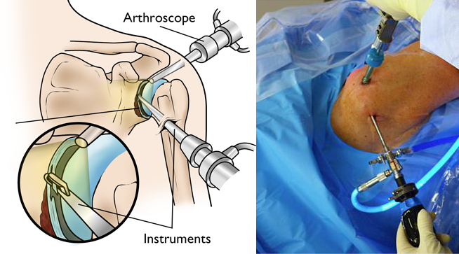
SLAP tears are typically treated with arthroscopic surgery.
- If the tear is limited to the labrum and does not involve the biceps tendon attachment, your surgeon will often remove the torn part of the labrum and correct any other related problems while leaving the biceps tendon in place.
- If the tear extends into the biceps tendon or if the tendon is detached, the biceps tendon may also need treatment. Your surgeon may decide either to repair the tendon, release the tendon, or transfer the tendon to a safer location using absorbable tacks, anchors, screws. Studies have shown that all of these techniques produce similar outcomes and there is not one that works best for all patients. Your surgeon will typically talk to you about which technique they recommend for you.
Bankart Lesion (Anteroinferior Labral Tear) or Posterior Labral Tear Treatment
- Tears of the labrum located in the bottom half of the socket may result in shoulder instability (Bankart lesion) or pain (posterior labral tear). In these cases, the surgeon will typically reattach the labrum and ligaments with suture anchors. Sometimes, they will also tighten the shoulder socket tissue by folding it over and "pleating" it. This procedure may be done arthroscopically, or it may be an open procedure with a small incision on the front of the shoulder.
- If your injury is more severe, with fractures or bone loss, your surgeon may recommend a surgery that includes replacing lost bone with bone from either other parts of your shoulder or a donor. This will help make your shoulder stable again.
It should be noted that not all labral tears in the shoulder can be made better with surgery. This is especially true for labral tears caused by normal wear and tear of the joint, or osteoarthritis.
If your labral tear does not necessarily need to be treated with labral repair, your doctor will talk to you about nonsurgical treatment options or other surgeries that are typically used to treat osteoarthritis. More information about shoulder arthritis treatment can be found in the article Arthritis of the Shoulder.
Rehabilitation After Surgery
- After surgery, your surgeon will typically recommend that you wear a sling for 2 to 6 weeks, depending on the type of repair that was done.
- Your surgeon will also typically prescribe gentle, passive, pain-free range-of-motion exercises early on.
- After several weeks, your surgeon will prescribe further exercises to help you regain shoulder motion and strength. You may be sent to a physical therapist, who will guide you in these exercises.
- Your surgeon will likely recommend some restrictions, such as not lifting anything more than 5 pounds, for up to 12 weeks after surgery.
- Athletes can usually begin doing sport-specific exercises 12 weeks after surgery.
- The shoulder should be fully healed in 4 to 6 months.
Surgical Outcomes
Results from surgery are typically very good. Most patients will ultimately regain most of their strength and range of motion and will be able to return to their normal activities.

Contributed and/or Updated by
Peer-Reviewed by
AAOS does not endorse any treatments, procedures, products, or physicians referenced herein. This information is provided as an educational service and is not intended to serve as medical advice. Anyone seeking specific orthopaedic advice or assistance should consult his or her orthopaedic surgeon, or locate one in your area through the AAOS Find an Orthopaedist program on this website.
The content on this website may have been translated from English into other languages using artificial intelligence tools. The translations are provided for the convenience of readers and may not been reviewed or verified by medical professionals. AAOS makes no guarantees regarding the accuracy, reliability, or completeness of the translated information and disclaims all liability for any issues arising from its use. Users are strongly encouraged to refer to the original English content and consult qualified healthcare professionals before making any medical decisions. By using the translated content, you acknowledge and accept these limitations and assume full responsibility for any reliance on the information provided.